
 Checks
- Prenatal screening for Trisomy 21
- Non-invasive screening (Genatest®)
- Pregnancy calendar
- Specialized ultrasounds
IMPORTANT INFORMATION
During pregnancy monitoring visits, children are not allowed to be present during ultrasound exams or during prenatal consultations to avoid interfering with exams and to facilitate the mission of the medical and nursing teams. More infoThere is a daycare available that is open Monday to Friday, from 8:30 a.m. to 12:30 p.m. and from 1 p.m. to 5 p.m. for children from one year to 10 years of age.
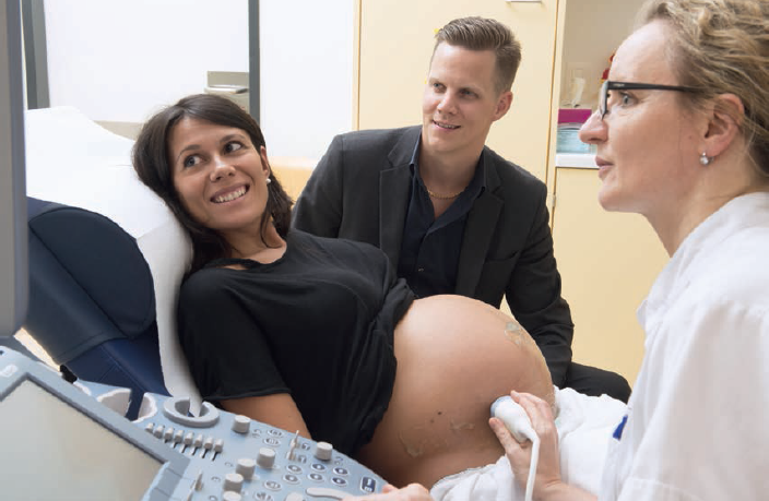
Checks
During the nine months, various checks are offered to rule out the risk of complications: clinical exams, weight measurements, blood pressure and uterine height checks, blood tests, urinalyses, auscultations of the baby’s heart, etc.
The ultrasound determines the term of the pregnancy and enables the exclusion of quite a few complications in the baby (malformations, growth problems). At the end of the pregnancy, if necessary, it can be used to estimate the baby’s size.
The uterine height is used to evaluate the growth of the fetus, the development of the uterus, and the quantity of amniotic fluid. A measuring tape is used to measure uterine height.
Prenatal screening for Trisomy 21
The first trimester screening test is offered to all pregnant women, but it is not mandatory. It is used to estimate your risk that the fetus is carrying a genetic abnormality, such as Trisomy 21 (or Down syndrome). This calculation of statistical probability is the result of a link between three parameters: your age, the presence of certain markers in your blood and the nuchal translucency, and the ultrasound scan of the thickness of the baby’s neck (between the 11th and 14th week of pregnancy). Depending on the level of risk, other diagnostic tests are offered (usually with a risk greater than 1 in 1000).
Non-invasive screening (Genatest®)
This test is offered starting in the 11th week of pregnancy to screen for Trisomy 21 (and Trisomy 18 and 13) in the fetus, if the prenatal screening shows a result greater than 1 in 1000. It is carried out by means of a blood test, and involves the analysis of the genetic material of the fetus released by the placenta into your blood. It involves no risk of danger to the unborn child. If the result is abnormal (strong suspicion of Trisomy), this must be confirmed by an invasive diagnostic exam (choriocentesis or amniocentesis).
The cost for this test is covered by health insurance for pregnant women whose fetus presents a moderate to high level of risk of Trisomy at the end of the first trimester screening test. However, any pregnant woman who wishes to could have this test performed, but the cost for it will not be reimbursed if the risk is not greater than 1 in 1000.
Pregnancy calendar
Download the pregnancy calendar
Specialized ultrasounds
Specialized ultrasounds are performed as part of specialized consultations in fetal medicine:
- Pregnancy consultation in case of maternal or fetal risks
- Fetal cardiology consultation
- Consultation in case of multiple pregnancies
They are carried out in multidisciplinary collaboration with the High Risk Obstetrics Unit and various partner divisions.
Fetal Medicine/Pathology consultation
Recommended monitoring of a normal pregnancy consists of a screening test in the first trimester, a morphology test in the second trimester and possibly a growth check in thethird trimester.
In most cases, these exams show normal findings. During a pregnancy ultrasound exam, the exam may show a fetal problem that requires treatment or special care. During the fetal medicine consultation, couples can meet the obstetrician, geneticist, and pediatrician, depending on the pathology presented by the fetus, to discuss diagnostic and therapeutic options that are tailored to the situation.
This consultation is also offered to all couples who have experienced a serious fetal pathology that resulted in a medical termination of the pregnancy. It usually takes place six weeks after hospitalization.
Cardio-pediatrics
The obstetrician sometimes consults with the pediatrician specializing in cardiologyin case of a medical antecedent or maternal pathology that is likely to influence morphology or fetal cardiac function (e.g. insulin-dependent diabetes, anti-epileptic treatment, an antecedent of maternal or family cardiac malformation), or when the cardiac examination of the fetus is considered to be abnormal or difficult during a prenatal ultrasound exam.
Consultation for twins
Twin pregnancies are specially monitored with an ultrasound follow-up plan and clinical follow-up because of a greater potential for specific complications with multiple pregnancies. The follow-up schedule is therefore different from single pregnancies. At each consultation, we check to ensure that the development of the pregnancy is still going well and look for potential complications.
Fetal sampling
Fetal sampling mostly consists of amniotic fluid samples (amniocentesis) or chorionic villi samples (choriocentesis) in order to determine the fetal karyotype. Choriocentesis is performed between 11 and 13 weeks of amenorrhea, while amniocentesis is performed starting at 15 weeks of amenorrhea. Amniocentesis has been routinely performed for many years under ultrasound control, starting at 15 weeks of amenorrhea. Insertion of a needle into the uterus enables us to take an amniotic fluid sample and study of fetal cells in it. This technique is not risk-free and leads to an involuntary abortion in 1% of cases.
Choriocentesis, also called “chorionic villi sampling”, consists of a biopsy of the placenta between 11 and 13 weeks of amenorrhea using a transabdominal or transcervical method. It enables an earlier diagnosis of certain chromosomal abnormalities and genetic diseases (Medical Genetics Division). It also involves a 1 to 2% risk of abortion.
Fetal interventions and fetoscopy
The Fetal medicine and ultrasonography unit is currently developing a new facet in obstetric activity through the implementation of interventional procedures, such as fetoscopy or use of a laser. These samples also make it possible to look for genetic diseases or infectious agents. The risk of complications from these procedures is 1% of cases . In 0.5% cases, membrane rupture-type complications and hematoma bleeding occur, and in the remaining 0.5% cases, involuntary abortions occur.
Umbilical cord blood sampling (cordocentesis) can be performed to analyze the fetal blood for the impact of various diseases. Access to the umbilical cord also allows intrauterine treatment, for example, fetal transfusions, which are useful in case of anemia on Rh-alloimmunization, other types of anti-erythrocyte antibodies or infectious-origin antibodies (Parvovirus). However, fetal blood sampling is performed less frequently because of a few indications that require a cordocentesis and the possibility of obtaining the necessary information through amniocentesis (it was previously accessible only through cordocentesis).